Error message
Deprecated function: implode(): Passing glue string after array is deprecated. Swap the parameters in drupal_get_feeds() (line 394 of /home3/pathiipo/public_html/includes/common.inc).
With the advent of whole slide digital scanners, histopathology slides can be digitized into very high-resolution digital images, realizing a new “big data” stream that can potentially rival “omics data” in size and complexity. Just as with the analysis of high-throughput genetic and expression data, the application of sophisticated image analytic tools and data pipelines can render the often passive data of digital pathology (DP) archives into a powerful source for:
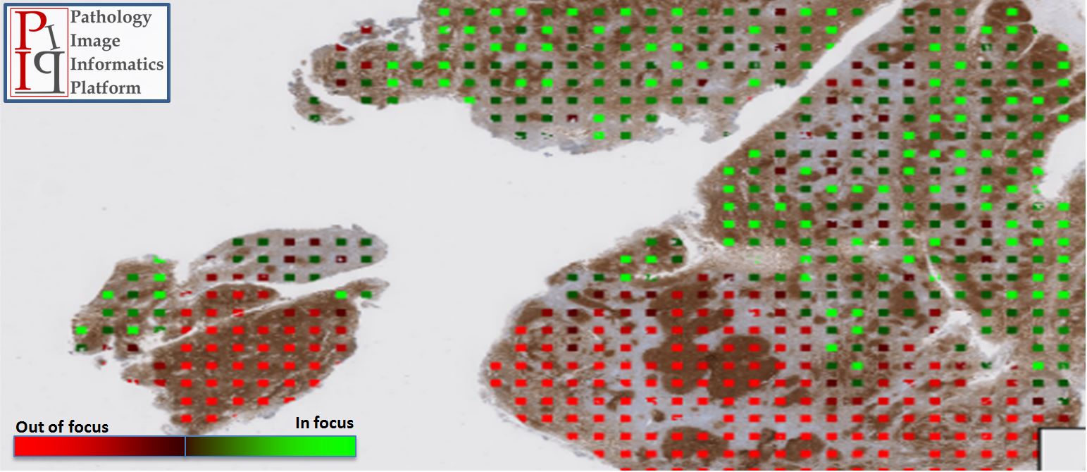
Precise focusing is one of the important parameters to obtain high-quality images. Unfortunately, whole slide images produced by commercial scanners may result in out-of-focus areas. The main reason for the blurred images is the improper focusing on the tissue. Although the scanners are able to calculate the precise focusing parameter for a specific point, the regions between two focus points can be out-of-focus due to the 3d structure of the tissue.
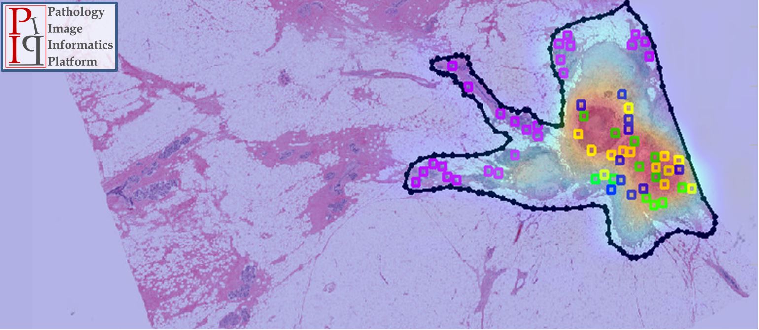
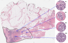
Neoadjuvant systemic therapy (NAT) for breast cancer (BC) is used to treat locally advanced BC prior to breast-conserving surgery. The response of cancer to NAT provides an opportunity to monitor the effectiveness of therapy, and a pathological complete response has been shown to predict survival and local recurrence. More recently a quantitative measure of response, the RCB, has been shown to provide valuable prognostic information but it is time-consuming to evaluate.
Tuesday 13 February 2018, 5:00 PM - 7:00 PM, Salon D/E
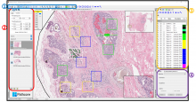
Sedeen: An extensible viewer for digital pathology
Anne Martel, Sunnybrook Research Institute, Toronto, Metin Gurcan, Wake Forest, NC, Anant Madabhushi, Case Western Reserve University, OH
Location:
Salon D/E, Marriott Marquis Houston Houston, Texas, United States
Event Date:
Tuesday, February 13, 2018
Our paper describing the PIIP has just been published in a special issue of Cancer Research.
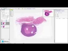
This video demonstrates how to align and overlay two images using the Sedeen viewer. The Sedeen Viewer can be downloaded from the Pathcore website http://pathcore.ca/sedeen/
MICCAI 2016, the 19th International Conference on Medical Image Computing and Computer Assisted Intervention, will be held from October 17th to 21st, 2016 in Athens, Greece. MICCAI 2016 is organized in collaboration with Bogazici, Sabanci, and Istanbul Technical Universities.
Workshop on Radio-Path-Omics: Analytic Tools for Correlating, Co-registering and Combining Radiologic, Pathologic, and Molecular Data
Oct 21, 2016 11:00 – 13:00
Athenaeum CC II-III
Event Date:
Monday, October 17, 2016 to Friday, October 21, 2016
SPIE Medical Imaging conference in San Diego
Event Date:
Wednesday, March 2, 2016 to Thursday, March 3, 2016