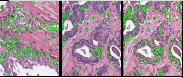
Pathology Stain Normalization
Owing the differences in slide preparation and staining protocols across labs, some level of color standardization may be necessary for initial detection, segmentation of histological structures. The contact PI’s group has previously developed effective color standardization methods. However, the specific algorithm that will be integrated within PIIP will be an Expectation Maximization (EM)-based color standardization scheme (developed by the contact PI) that improves color reproducibility in pathology by independently aligning color distributions from different tissue classes (e.g. epithelium, stroma).
The PIIP uses an architecture that is specifically designed to facilitate algorithm pipelining. Color and stain normalization apps will be developed as plugin modules in the PIIP using the filter ideology. Filters are a unique set of algorithms that generate modified images rather than specific displayable outputs like contours or segmentation masks and perform vital preprocessing operations for image analysis algorithms. Filter outputs, images in their own right, can then become inputs to other image segmentation or registration algorithms.
